 A dental cone beam CT is a kind of X-Ray used when we need to see more detail than a regular dental X-Ray can provide. Our cone beam CT uses technology that can allow it to create a 3D image of your dental structures, soft tissues, nerve paths, and facial bones in one single scan. These more detailed images will allow our skilled endodontists to more accurately create your treatment plan.
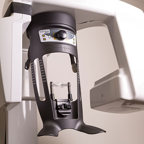
What to Expect During a Cone Beam CT Scan
Before receiving your cone beam CT scan, you will be asked to remove any metal objects including jewelry, bobby pins, dentures, eyeglasses, or bras with a metal underwire. This is because metal objects can affect the images generated by the cone beam CT.
During the scan, an X-Ray beam will move around your head, producing many views and images as it does so. This beam is cone-shaped, which is what gives the machine its name. These scans will be very detailed and high-quality. Additionally, a cone beam CT produces much less radiation compared to a conventional CT scan.
Reasons for Cone Beam CT
In endodontics, cone beam CT images are used to create treatment plans as well as view the canals of teeth that have become calcified. Cone beam CT scans allow us to navigate within the root canal system of the teeth and can show any abnormal anatomy of the tooth or the internal canals.
If you have questions or concerns about our cone beam CT technology, please don’t hesitate to give us a call. Syrpes & Pangborn is located here in Centennial, Colorado.
 Our Doctors
Here at Syrpes & Pangborn, we strive to offer a standard of excellence in personalized endodontic care. New Patients
Our office is open Monday - Friday from 7:00am - 4:30pm. We will schedule your appointment as promptly as possible. Root Canal
The main goal of a root canal is to save the natural tooth. Our doctors will help restore the tooth to its full function.